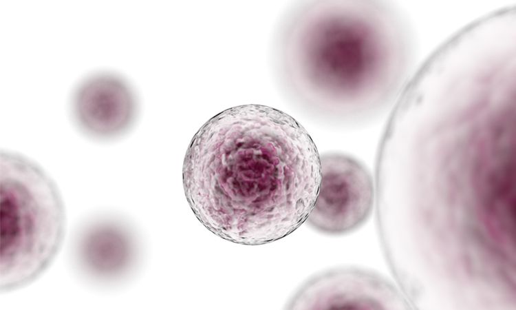
What are senescent cells?
Senescent cells are cells that can no longer divide or support the tissue they are part of (7, 13). This is a permanent state of cell cycle arrest brought on by cellular stress (4). A senescent cell is in its twilight: not quite dead, but not functioning as it had originally (14).
One of the main theories for cellular senescence has to do with telomerase. Telomerase is the “end cap” of DNA that protects DNA loss through cell division, although these end caps shrink with every cell division (3, 15). After a certain number of divisions, the telomeres become too short and the cell then becomes senescent (3, 15).
These senescent cells linger in the body and do not die because they underproduce the protein that triggers programmed cell death or apoptosis (8). The older you get the more senescent cells accumulate in the body and potentially lead to age-related complications (8).
Senescent Cells and Aging
Cellular senescence is one of the various processes that lead to aging (8). As you grow older, an increasing number of cells enter into this senescent state. In this state, they excrete damaging chemical signals that cause inflammation and drive the aging process (8). This includes pro-inflammatory proteins, cytokines, and signals, known as the senescence-associated secretory phenotype (SASP). SASP blocks a variety of important cellular processes prevents stem cells from effectively repairing damaged tissue, and is associated with the development of age-related diseases (5, 8).
This combination excreted from senescent cells can also encourage other nearby healthy cells to become senescent, leading to a downward spiral of increasingly poor tissue repair (8, 14). This means a small number of senescent cells can have a dramatic effect on the surrounding tissue, interfering with tissue homeostasis and regeneration (5).
Although many cells do die on their own, all somatic cells (non-reproductive cells) that divide have the ability to undergo senescence (14).
Is senescence always bad?
In young, healthy tissue, SASP secretions are said to be part of a restorative process, where damaged cells stimulate repair in nearby tissues by sending out a distress signal to the immune system to eliminate them (14). In this case, senescence is generally speaking a good thing as it keeps us safe from damaged cells reproducing while promoting healthy tissue repair. Although at some point, senescent cells begin to accumulate (5).
Clearing away unwanted senescent cells is the job of the immune system, and replacing the lost cells requires stem cells to repopulate the tissue (8). As we age, the ability of the immune system to clear these cells begins to fail, leading to this accumulation (4, 5). Senescent cell accumulation then increases inflammation, reducing the ability of stem cells to replace loses which begins the downward spiral where senescence is no longer a safety checkpoint and becomes a true driver of the aging process (4).
What does senescent cell removal – or clearance – result in?
After this realization, many have studied the possibility of removing these senescent cells through senolytic therapies (8). Past studies have tested potential senolytic therapies on animals, with some showing an increase in the healthy period of life and even maximum lifespan. Other studies have shown that senolytics have the potential to treat age-related diseases (14).
In some studies, just removing thirty percent of senescent cells showed significant improvements in age-related decline (8). This suggests the possibility of selectively removing senescent cells using senolytics to alleviate the symptoms of aging and also promote general health as we age (8, 14).
Other experiments have suggested that senescent cells accumulate in aging organs and that eliminating them could help alleviate certain illnesses (14). A 2016 study, also suggested extened lifespan of normally aging mice (14). Studies such as these suggest that just removing senescent cells could stimulate new tissue production, and jump-start some of the tissue’s natural repair mechanisms.
Are all senescent cells the same?
Surprisingly, senescent cells are all slightly different in each tissue. They secrete different cytokines, express different extracellular proteins, and use different tactics to avoid death (14). This has made it a challenge for labs to detect and visualize senescent cells as a whole. “There is nothing definitive about a senescent cell. Nothing. Period” says one researcher (14).
Therefore targeting the different tissues would require different types of senolytics.
Should we then look to wipe out senescent cells completely?
As the previously mentioned studies displayed, no it is not necessary to completely eradicate all senescent cells (14). Just getting rid of most of them is enough to make a difference (5).
Also, senolytic drugs clear only senescent cells that are already present—they won’t prevent the formation of more senescent cells in the future. This means that senescence can continue to perform its original role in the body, but while managing the negative effects of senescent cell buildup (5, 14).
How does it apply to exercise?
Our most recent Senactiv® human clinical study has concluded a decrease in senescent cells in active muscles. This means the muscles (the largest tissue system in the body) should be able to perform at a “younger” level due to this clearing away of senescent cells. It is an anti-aging remedy for the muscle, promoting new healthy cells in the area, and potentially decreasing inflammation from the proinflammatory cytokine secretion from old senescent cell buildup.
Decrease in Apoptotic Cells in Muscle Post Exercise to Aid Recovery
What is apoptosis?
Apoptosis, or programmed cell death, is the body’s natural way of clearing away old cells to make room for newly formed cells (1, 6). Apoptosis is also responsible for eliminating potentially hazardous cells, and maintaining balance within the body (1). Cells within the body are constantly being destroyed and replaced in a constant cycle.
When a cell is compelled to “die”, proteins called caspases are activated to carry out the task (6). They break down the apoptotic cells in an organized and controllable manner, which is then cleaned up by macrophages, the cleaning crew of the tissue (1, 6).
That’s not to say that apoptosis is a perfect process. Sometimes, the wrong cells kill themselves due to other factors including DNA damage, and other times the ones that should be destroyed linger in the body (6).
Process Benefits
Apoptosis is essential to human development and healthy survival. As our brains develop, the body creates millions of cells than it needs; the ones that don’t form synaptic connections undergo apoptosis so that the remaining cells can function properly (1, 6). Also, as a fetus develops and grows, it has webbed hands due to its aqueous environment. Apoptosis is the reason individual fingers develop the way they do. The unneeded cells creating the webbing are destroyed, creating the individual fingers we have when we are born (1).
On a more regular basis, apoptosis allows the body to clear out weak and unfit cells and allows healthy new cells to take their place (1, 6). When these cells stick around after their programmed death was supposed to occur, it can lead to the abnormal buildup of cells. Apoptosis involves the death of a cell, but it benefits the organism as a whole by eliminating potential bad cells and making room for newly formed cells into the body (1).
How does it relate to exercise?
Prolonged physical activity can cause skeletal muscle damage, with eccentric activity (lengthening contractions) and “large range of motion” activities (13). Previously it was believed that the damage was largely due to inflammatory and necrotic processes (which is an unplanned cell death that leads to inflammation), but recent evidence indicates an important role for apoptosis in adult muscle fibers during and after eccentric exercise (1, 13).
Although it remains unclear how and why apoptosis is induced in adult skeletal muscle after exercise, there are many plausible hypotheses that warrant further investigation (13). One of the leading hypotheses is that during exercise, muscle metabolism is increased, which leads to oxidative stress. Significant amounts of oxidants can lead to DNA damage which directly induces apoptosis. Type I muscle fibers, which are predominately oxidative, may be overwhelmed and unable to control the increased oxidative stress (13).
Additionally, the stress of the exercise increases catecholamine levels, which promote the induction of apoptosis (13). This added to the increase in oxidative stress could then signal the cell to undergo apoptosis. Apoptotic cell death induced by exercise in tissues exposed to specific stresses may be a normal process used to remove partially damaged cells (13). Excessive and/or eccentric exercise may cause significant mechanical damage, followed by an inflammatory response, leading to necrosis and apoptosis (13).
What does this mean for Senactiv®?
The decrease in apoptotic cells in this instance is due to the activation of phagocytosis in macrophages responsible for clearing away the unfit or broken-down cells within the body. The recent Senactiv® human clinical study states that even though exercise generally increases the movement of white blood cells and “cleans up” cells into the muscle, it does not always activate phagocytosis to clear away the broken-down apoptotic cells and senescent cells (16). With Senactiv® phagocytosis activation, it is able to clear these types of cells away leading to a potential decrease in both apoptotic and senescent cells in active muscles.
Reduces Leukocyte Infiltration in Exercising Muscles
What is a leukocyte?
Leukocytes, or white blood cells, are a vital part of the body’s immune system. These cells help to fight infection and counteract foreign substances and disease (11). Blood tests often look for white blood cell count and location to check for conditions such as infection, inflammation, allergies, and leukemia (11). In this case, muscle inflammation was our point of the study.
What is Infiltration?
Briefly defined, Infiltration is the diffusion or accumulation (in tissue) of foreign substances in amounts in excess of the normal. The material collected in those tissues or cells is called infiltrate (2).
Leukocyte Infiltration?
From these definitions, Leukocyte infiltration is the accumulation of white blood cells in muscle tissue.
Why would there be an accumulation?
Eccentric muscle contractions induce muscle damage that results in the acute phase response. This is where the body recognizes the problem, reacts with cell activation and cytokine release, then deals with the problem through leukocyte infiltration and phagocytosis, resolving the inflammation problem in the damaged tissue (7).
The accumulation of these inflammatory cells (leukocytes) in the muscle tissue is a sign of this exercise-induced muscle damage. Leukocyte accumulation in muscles has had relatively consistent findings as a response to moderate to severe muscle damage, typically induced by maximal eccentric exercise across a large range of motion (12).
How does it relate to exercise?
Therefore, a result of reduced leukocyte infiltration provides further proof of reduced inflammation of the muscle, as well as reduced muscle damage from extreme exercise.
Activates Macrophage in Exercising Muscles
What are macrophages?
Macrophages are a specialized type of white blood cell that is involved in the detection, and destruction of bacteria, microscopic particles, dead cells, and other harmful organisms (9, 10).
Macrophages develop from white blood cells called monocytes, which are produced by stem cells in our bone marrow. Monocytes move through the bloodstream and mature into macrophages once they leave the blood (10). They live for months, patrolling our cells and organs to keep them clean, basically acting as cellular super-janitors (10).
Why are they important?
Macrophages are a very important part of our immune system. The word ‘macrophage’ literally means ‘big eater.’ They are amoeba-like organisms that clean our bodies of microscopic debris and invaders. They have the ability to locate and engulfing or “eating” particles, such as bacteria, viruses, fungi, and parasites to protect the rest of the tissue from damage (9, 10).
Macrophages use a process called phagocytosis to destroy and get rid of unwanted particles in the body. Phagocytosis literally means ‘eat cell.’ First, the macrophage engulfs the particle, then a pocket called a phagosome is formed around it. Enzymes are released into the pocket to help digest the particle, then the remaining debris, or what is left of the particle, exits the macrophage to be absorbed back into the body (9, 10).
How does it relate to exercise?
This is a vital process involved in many of the above-mentioned tasks. Phagocytosis and macrophage activation are involved in the removal of senescent cells and the clearing away of broken-down apoptotic cells. This process also helps to reduce inflammation by “eating” inflammatory substances and other harm-causing particles. According to the recent Senactiv® human clinical study, macrophage activation is essential for exercise-induced senescent cell clearance in exercising skeletal muscle (16).
References
- Apoptosis. (n.d.). Retrieved from https://www.khanacademy.org/science/biology/developmental-biology/apoptosis-in-development/a/apoptosis
- Belkin, M., Lamorte, W. L., Wright, J. G., & Hobson, R. W. (1989). The role of leukocytes in the pathophysiology of skeletal muscle ischemic injury. Journal of Vascular Surgery,10(1), 0014-0019. doi:10.1067/mva.1989.vs0100014
- Bernadotte, A., Mikhelson, V. M., & Spivak, I. M. (2016). Markers of cellular senescence. Telomere shortening as a marker of cellular senescence. Aging,8(1), 3-11. doi:10.18632/aging.100871
- Childs, B. G., Gluscevic, M., Baker, D. J., Laberge, R., Marquess, D., Dananberg, J., & Deursen, J. M. (2017). Senescent cells: An emerging target for diseases of ageing. Nature Reviews Drug Discovery, 16(10), 718-735. doi:10.1038/nrd.2017.116
- Deursen, J. M. (2014, May 22). The role of senescent cells in ageing. Retrieved from https://www.ncbi.nlm.nih.gov/pmc/articles/PMC4214092/>
- Edmonds, M. (2018, March 08). What is apoptosis? Retrieved from https://science.howstuffworks.com/life/cellular-microscopic/apoptosis.htm
- Hamada, K., Vannier, E., Sacheck, J. M., Witsell, A. L., & Roubenoff, R. (2005). Senescence of human skeletal muscle impairs the local inflammatory cytokine response to acute eccentric exercise. The FASEB Journal,19(2), 264-266. doi:10.1096/fj.03-1286fje
- Hill, S. (2017, March 25). Hallmark of Aging Reversed by Senescent Cell Removal |. Retrieved from https://www.leafscience.org/senescent-cells
- Macrophages. (n.d.). Retrieved from https://www.immunology.org/public-information/bitesized-immunology/cells/macrophages
- McDougal, W. (n.d.). Macrophages: Definition, Function & Types. Retrieved from https://study.com/academy/lesson/macrophages-definition-function-types.html
- NCI Dictionary of Cancer Terms. (n.d.). Retrieved from https://www.cancer.gov/publications/dictionaries/cancer-terms/def/leukocyte
- Peake, J. M., Neubauer, O., Della Gatta, P. A., & Nosaka, K. (n.d.). Muscle damage and inflammation during recovery from exercise. Manuscript, Institute of Health and Biomedical Innovation, Queensland University of Technology, Brisbane, Australia.
- PHANEUF, S., & LEEUWENBURGH, C. (n.d.). Symposium: Antioxidant and Redox Regulation of Cellular Signaling. Manuscript, Apoptosis and exercise, Biochemistry of Aging Laboratory, Center for Exercise Science, College of Health and Human Performance, University of Florida, Gainesville, FL 32611.
- Scudellari, M. (2017, October 25). To Stay Young, Kill Zombie Cells. Retrieved from https://www.scientificamerican.com/article/to-stay-young-kill-zombie-cells/
- Telomerase Enzyme | Telomere Shortening | TA 65 Supplement. (n.d.). Retrieved from https://www.tasciences.com/telomeres-and-cellular-aging/
- Wu, J., Saovieng, S., Cheng, I., Liu, T., Hong, S., Lin, C., . . . Kuo, C. (2018). Ginsenoside Rg1 supplementation clears senescence-associated β-galactosidase in exercising human skeletal muscle. Journal of Ginseng Research. doi:10.1016/j.jgr.2018.06.002